Welcome to Cruciate-Ligament.com!
This site was created to assist dog owners in recognizing potential problems with their dog’s cruciate ligament.
This guide does not replace veterinary care or treatment. If you suspect that your dog is suffering, please visit your veterinarian immediately for a correct diagnosis.
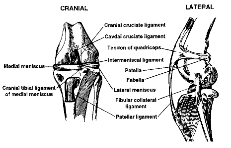
The knee is a joint that is formed by three bones:
- Femur (the long bone extending down from the hip)
- Tibia (the bone between the knee and ankle)
- Patella (the kneecap)
These bones are joined together by a number of ligaments, which are tough fibrous bands of tissue. Two ligaments crisscross in the joint from the femur to the tibia and are called cruciate ligaments.
The one towards the front of the leg is called the anterior cruciate ligament and the one crossing behind it is the posterior cruciate ligament. These ligaments prevent the ends of the femur and tibia from moving back and forth across each other.
What is the Cruciate Ligament?
The knee is a fairly complicated joint. It consists of the femur above, the tibia below, the kneecap (or “patella”) in front, and the bean-like fabellae behind. Chunks of cartilage called the medial and lateral menisci fit between the femur and tibia like cushions and there are an assortment of ligaments holding everything together allowing the knee to bend the way it should and keep it from bending the way it shouldn’t.
There are two cruciate ligaments which cross inside the knee joint: the anterior (or, more correctly in animals, cranial cruciate) and the posterior (or, more correctly in animals, the caudal cruciate). They are named for the side of the knee (front or back) where their lower attachment is found. The anterior cruciate prevents the tibia from slipping forward out from under the femur.
Symptoms of a Ruptured Ligament
Dogs who have ruptured their cruciate ligament will appear suddenly lame, and usually hold the foot of the affected leg off the ground.
The knee may become swollen. In time, the dog may start to use the leg again, but often lameness returns.
Want to learn how to save on the treatment of cruciate ligament problems? Click here
Want to check pricing and try our veterinary discount program, risk free? Click here
Which Dogs Are More at Risk?
Cruciate ligament damage is most commonly seen in overweight middle-aged dogs. Another group of dogs that suffer from this disease are those receiving corticosteroid medications. The problem appears suddenly – often after some sudden twisting movement or jumping up or out of an elevated location. The pet often yelps at the sudden pain that occurs.
In light to moderate weight breeds (less than 25 pounds), use of the knee often becomes normal with time and rest. Lightweight dogs are sometimes placed on anti-inflammatory and pain management medications as well as glucosamine and plan a program of rest and physical therapy while the joint heals.
A recent study identified the following breeds as being particularly at risk for this phenomenon: Neapolitan mastiff, Newfoundland, Akita, St. Bernard, Rottweiler, Chesapeake Bay retriever, and American Staffordshire terrier.
How Do Ruptures Happen?
A torn cruciate ligament can occur in any dog if just the wrong forces impact the knee joint. Most commonly seen in larger breeds of dogs and in dogs that are overweight. Obesity puts too much weight on the knee and overweight dogs tend to have more occurrences of ruptured cruciate ligaments.
There are several clinical pictures seen with ruptured cruciates. One is a young athletic dog playing roughly who takes a bad step and injures the knee while playing. This is usually a very sudden lameness in a young large breed dog.
When the anterior cruciate ligament ruptures (is torn), the joint becomes unstable and the femur and tibia can move back and forth across each other. The anterior cruciate ligament is most commonly torn when the dog twists on his hind leg. The twisting motion puts too much tension on the ligament and it tears. This often occurs if the dog slips on a slippery surface, makes a sudden turn while running, or is hit by a car.
Want to learn how to save on the treatment of cruciate ligament problems? Click here
Want to check pricing and try our veterinary discount program, risk free? Click here
Veterinarian Diagnosis
The veterinarian stabilizes the position of the femur with one hand and manipulates the tibia with the other hand. If the tibia moves forward (like a drawer being opened), the cruciate ligament is ruptured.
Another test that can be used is the “Tibial Compression test” where the veterinarian stabilizes the femur with one hand and flexes the ankle with the other hand. If the ligament is ruptured, again the tibia moves abnormally forward.
If the rupture occurred some time ago, there will be swelling on side of the knee joint that faces the other leg. This is called a “medial buttress” and is a sign that arthritis is well along. It is not unusual for animals to be tense or frightened at the vet’s office. Tense muscles can temporarily stabilize the knee preventing demonstration of the drawer sign during examination. Often sedation is needed to get a good evaluation of the knee. This is especially true with larger dogs.
Since arthritis can set in relatively quickly after a cruciate ligament rupture, radiographs to assess arthritis are a good idea. Another reason for radiographs is that occasionally when the cruciate ligament tears, a piece of bone where the ligament attaches to the tibia breaks off as well. This will require repair and the surgeon will need to know about it before beginning surgery.
Is It Too Late?
Without an intact cruciate ligament, the knee is unstable. Wear between the bones and meniscal cartilage becomes abnormal and the joint begins to develop degenerative changes. Bone spurs called “osteophytes” develop and chronic pain and loss of joint motion result. This process can be arrested by surgery but cannot be reversed.
- Osteophytes are evident as soon as 1-3 weeks after the rupture in some patients
- In one study a group of dogs was studied for 6 months after cruciate rupture. At the end of 6 months, 85% of dogs under 30 lbs of body weight had regained near normal or improved function while only 19% of dogs over 30lbs had regained near normal function. Both groups of dogs required at least 4 months to show maximum improvement.
A dog with arthritis pain from an old cruciate rupture may still benefit from a TPLO surgery. It may be worth having a surgery specialist take a look at the knee. Most cases must make do with medical management.
If the rupture occurred some time ago, there will be swelling on side of the knee joint that faces the other leg. This is called a “medial buttress” and is a sign that arthritis is well along. It is not unusual for animals to be tense or frightened at the vet’s office. Tense muscles can temporarily stabilize the knee preventing demonstration of the drawer sign during examination. Often sedation is needed to get a good evaluation of the knee. This is especially true with larger dogs.
Since arthritis can set in relatively quickly after a cruciate ligament rupture, radiographs to assess arthritis are a good idea. Another reason for radiographs is that occasionally when the cruciate ligament tears, a piece of bone where the ligament attaches to the tibia breaks off as well. This will require repair and the surgeon will need to know about it before beginning surgery.
Want to learn how to save on the treatment of cruciate ligament problems? Click here
Want to check pricing and try our veterinary discount program, risk free? Click here
Surgery
There are 3 different surgical repair techniques commonly used:
EXTRACAPSULAR REPAIR:
lateral orthopedic wire suture in a dog’s left stifle (knee)
This procedure is currently favored as it can be performed in a relatively shorter surgery time than the other procedures. The knee joint is opened and inspected. The torn or partly torn cruciate ligament is removed. Any bone spurs of significant size are bitten away with an instrument called a “rongeur.” If the meniscus is torn, the damaged portion is removed. A wire or very large, strong suture is passed around the fabella behind the knee and through a hole drilled in the front of the tibia. This tightens the joint to prevent the drawer motion, effectively taking over the job of the cruciate ligament.
INTRACAPSULAR REPAIR:
This procedure has fallen out of favor lately as it has been unable to demonstrate superior results to the extracapsular technique described above. Intracapsular repair intuitively seems like it should do better as it uses living tissue (rather than an artificial material) to essentially make a new ligament. This takes more time surgically. As with the extracapsular repair, the knee joint is opened, fragments of the ligament are removed, as is damaged meniscus. After this a strip of connective tissue is dissected locally and passed through the middle of the joint exactly where the cruciate ligament used to be. The “new ligament” is attached at the opposite end to an implant or simply sewn into place.
TIBIAL PLATEAU LEVELING OSTEOTOMY (TPLO)
This procedure uses a fresh approach to the biomechanics of the knee joint and is meant to address the lack of success seen with the above two techniques long term in larger dogs. With this surgery the tibia is cut and rotated in such a way that the natural weight-bearing of the dog actually stabilizes the knee joint. As before the knee joint still must be opened and damaged meniscus removed. The cruciate remnants may or may not be removed depending on the degree of damage.
The TPLO rotaes (as indicated by the arrow) the sloped tibial plateau until it is perpendicular to the line between the stifle and the hock joint centers
This surgery is complex and involves special training in this specific technique. Many radiographs are necessary to calculate the angle of the osteotomy (the cut in the tibia). At this time the TPLO is felt to be the best way to repair a cruciate rupture regardless of the size of the dog and is probably the only procedure to be considered for dogs over 50lbs. This surgery typically costs twice as much as the extracapsular method.
What to Expect after Surgery
In the 3 different surgical repair techniques commonly used:
EXTRACAPSULAR REPAIR:
- The dog may carry the leg up for a good 2 weeks after surgery but will increase knee use over the next 2 months eventually returning to normal.
- The dog will require 8 weeks of exercise restriction after surgery (no running, outside on a leash only including the backyard)
- The wire placed will break 2-12 months after surgery and the dog’s own healed tissue will “hold” the knee.
INTRACAPSULAR REPAIR:
- Bandaging for a couple of weeks after surgery is commonly recommended.
- Again, the dog may not bear weight for a good two weeks after surgery and will likely require 2 months to return to normal function.
- Again, 8 weeks of exercise restriction will be necessary for healing.
TIBIAL PLATEAU LEVELING OSTEOTOMY (TPLO)
- Most dogs are touching their toes to the ground by 10 days after surgery though it can take up to 3 weeks.
- As with other techniques 8 weeks of exercise restriction are needed.
- Full function is generally achieved 3-4 months after surgery and the dog may return to normal activity.
Long Term Results
Surgical methods usually have good results. When widely differing procedures all result in improvement with time it is wise to question whether time itself may be the curative element. None-the-less, it is considered good practice in veterinary medicine in the United States to treat all these cases in larger breeds surgically. Quite a few of these dogs will rupture the cruciate ligament of the remaining good leg within three years. Weight reduction helps keep the affected knee stable. So does avoiding future extreme activities for your pet.
Want to learn how to save on the treatment of cruciate ligament problems? Click here
Want to check pricing and try our veterinary discount program, risk free? Click here

1. Enroll your pet. It takes less than 2 minutes!

2. Take your ID card to a local network veterinarian

3. The vet gives you an instant discount on all in-house medical services
![]()
![]()
![]()
![]()